About this article
Medically reviewed- Written by
- Dr Paul Watson
- Medically reviewed by
- Dr Han Simpson
- Published
- April 8, 2026
- Medical review date
- September 8, 2026
- Evidence searched through
- September 8, 2026
- Next review due
- September 8, 2027
- Intended audience
- General Readers & Patients
- Geographic scope
- Worldwide
- Conflicts of interest
- None declared.
- Funding
- Website Owner
- Report a correction
- Report an error or editorial concern
- Medical disclaimer
- Read the medical disclaimer
A patient comes in with a fullness behind the knee. The referral says “rule out Baker’s cyst.” The ultrasound comes back and does exactly one thing: it says “lump behind knee not Baker’s cyst”.
That is the point where many workups drift. The common diagnosis has been excluded, but no one has yet replaced it with a disciplined alternative. In practice, that is where mistakes happen. Some masses are harmless and slow-growing. Others are vascular, thrombotic, infectious, or neoplastic. They can sit in the same small anatomical space and present with very similar symptoms.

A lump behind knee not baker’s cyst is not a diagnosis. It is a diagnostic fork in the road.

Introduction When a Knee Lump Is Not a Simple Cyst
Baker’s cysts are common enough that they become the default assumption. That is understandable. In adults, asymptomatic Baker’s cysts are reported in 4.7% to 37% of cases, and when they are symptomatic they are often tied to intra-articular pathology, with osteoarthritis accounting for 50.6% of symptomatic cases according to this open-access review of Baker’s cyst epidemiology and associated disorders.
That prevalence shapes clinical thinking. It also creates a trap.
When the first scan says “not a Baker’s cyst,” some clinicians relax too early if the patient looks otherwise well. Others overreact and order every test available. Neither approach is efficient. The better move is to reset the problem and ask a more useful question: which structure in the popliteal fossa is producing the mass-like finding?
Why the negative ultrasound matters
A negative result for Baker’s cyst narrows one branch of the tree. It does not clear the patient.
The mass could arise from a vessel, a lymph node, tendon-related tissue, subcutaneous fat, infected soft tissue, or a deeper soft tissue lesion. The practical challenge is that many of these possibilities overlap on history alone. Patients often describe “tightness,” “swelling,” “pressure,” or “a knot.” Those words do not separate a lipoma from a thrombosed vein or a solid tumor.
Clinical takeaway: Once a Baker’s cyst is excluded, stop thinking in labels and start thinking in compartments, tissue type, and urgency.
What changes in the next five minutes
The next steps are not exotic. They are structured.
You need to decide three things early:
- Is this vascular or thrombotic? A pulsatile mass or features suspicious for DVT change the urgency immediately.
- Is this inflammatory or infectious? Warmth, erythema, tenderness, and systemic illness push the workup in a different direction.
- Is this a solid mass that has not been characterized properly? That is where incomplete reassurance from first-line imaging causes trouble.
A good clinician does not need every answer at once. A good clinician needs to recognize what cannot be safely missed.
Mapping the Popliteal Fossa Anatomy
If you do not have a mental map of the popliteal fossa, the differential remains fuzzy. The easiest way to think about it is as a crowded passageway behind the knee where several systems cross a narrow space.
The structures are layered, and that matters because the character of the lump often reflects where it comes from.
The neighborhood you are examining
The vascular structures are the highest-stakes residents. The popliteal artery and vein traverse the fossa. A vascular lesion can produce a mass, but the exam may also reveal a bruit, altered distal perfusion, venous congestion, or tenderness with calf symptoms.
The nerves include the tibial nerve and the common fibular nerve as they pass near the region. These do not usually create a classic visible lump, but masses in this area can compress them and produce numbness, radiating discomfort, or weakness.
The musculotendinous structures include the hamstrings superiorly and the heads of gastrocnemius inferiorly. Thickening, tendon injury, or adjacent soft tissue change can be mistaken for a discrete popliteal mass, especially when the exam is done with the knee in only one position.
To ground that anatomy visually, this short review is useful:
Why lymphatic and soft tissue structures matter
The popliteal lymph nodes are easy to forget until they become the answer. They can enlarge with local infection, inflammatory disease, or systemic illness. On exam, they may present as tender or soft nodular lesions rather than the smooth fluid contour many people expect from a cyst.
The fat and soft tissue envelope also creates room for benign masses such as lipomas and for more concerning soft tissue tumors. These may feel deceptively superficial at first exam, particularly in larger patients or when swelling obscures depth.
Anatomy guides the first bedside distinction
When I teach this exam, I ask trainees to classify the lump before naming it:
- Pulsatile or non-pulsatile
- Soft and mobile, or firm and fixed
- Superficial or deep
- Focal nodule, or diffuse fullness
- Painful with warmth, or painless with pressure symptoms
That sequence does two things. It keeps the exam anchored in anatomy, and it prevents the common habit of deciding too early that every posterior knee lump must be a cyst.
The Broad Differential Diagnosis of a Popliteal Mass
Once Baker’s cyst is off the table, the differential becomes broad fast. The useful move is to group diagnoses by tissue and urgency, not by memorized lists.
A concise consumer-facing review still captures the key breadth well: a non-Baker’s popliteal lump can reflect lipomas, popliteal aneurysms, malignancies including osteosarcoma and sarcoma, abscesses, boils, lymphadenopathy, and deep vein thrombosis, among others, as summarized in this overview of causes of lumps behind the knee.
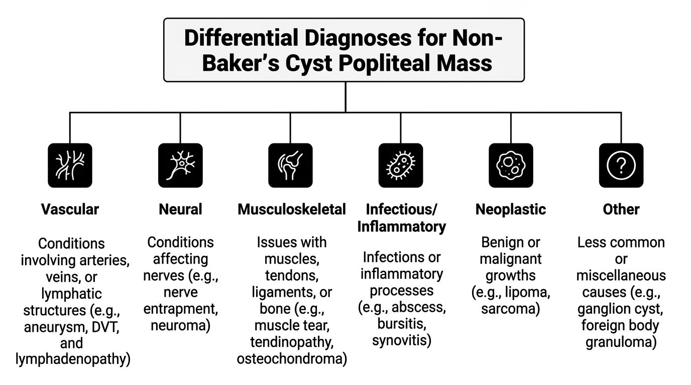
Vascular causes
These are the diagnoses you should actively try to exclude early.
A popliteal artery aneurysm may present as a posterior knee mass with pulsation or pressure symptoms. If the mass is mistaken for a benign cyst and compressed or aspirated, the error can be dangerous. The physical exam matters here. Feel for pulsation, assess distal pulses, and listen for a bruit when the story fits.
Deep vein thrombosis can also present as a mass-like fullness rather than the textbook picture of diffuse calf swelling. In the popliteal region, patients may localize the problem as “a lump behind the knee” because that is where they feel firmness or tenderness first.
Neoplastic causes
This category ranges from routine to high consequence.
A lipoma is usually soft, mobile, and relatively superficial. It often produces annoyance more than true pain. The danger is not the lipoma itself. The danger is assuming every soft tissue mass with slow growth is one.
A soft tissue sarcoma or other malignant lesion often behaves differently. Firmness, progressive enlargement, depth, and persistence despite a reassuring early scan should change your threshold for MRI and specialist referral. Bone-related malignancy such as osteosarcoma belongs in the broader differential when pain, bony symptoms, or concerning imaging findings suggest origin outside the soft tissue envelope.
Infectious and inflammatory causes
A posterior knee abscess can create a tender, warm, fluctuant or indurated mass. Patients may report fever, recent skin infection, or progressive local pain. The exam usually tells you this is not a quiet incidental lesion.
Lymphadenopathy in the popliteal region is less common than inguinal enlargement, which is one reason clinicians may overlook it. It may be tender in local infection or more indolent in systemic disease. Context matters: skin lesions, lower leg cellulitis, inflammatory disease, or generalized adenopathy should all be asked about.
Musculoskeletal and miscellaneous causes
Not every non-Baker’s popliteal finding is a true neoplasm or vascular lesion.
A ganglion-type cyst from tendon or adjacent soft tissue structures may mimic a popliteal cyst but arise from a different tissue plane. Tendon thickening, myofascial injury, and organized post-traumatic hematoma can also present as a palpable posterior fullness.
Bedside certainty tends to be overstated in this situation. If the tissue origin is not obvious, the right response is not confidence. It is better characterization.
A practical comparison at the bedside
| Condition | Typical Presentation | Key Feature(s) on Exam | Urgency |
|---|---|---|---|
| Lipoma | Slow-growing, often painless lump | Soft, mobile, superficial feel | Usually non-urgent |
| Popliteal artery aneurysm | Fullness behind knee, pressure symptoms | Pulsatile mass, possible bruit, vascular findings | Urgent vascular assessment |
| Deep vein thrombosis | Acute or subacute pain, swelling, tenderness | Firm tender fullness, warmth, calf symptoms | Urgent |
| Abscess or boil | Painful enlarging lump with inflammation | Warmth, erythema, tenderness, possible fluctuance | Prompt assessment |
| Lymphadenopathy | Nodular swelling, sometimes tender | Discrete node-like feel, associated local or systemic clues | Depends on cause |
| Soft tissue sarcoma | Persistent or enlarging deep mass | Firm, less mobile, may feel deep-seated | Urgent specialist workup |
| Ganglion or tendon-related lesion | Localized fullness with movement-related symptoms | Relation to tendon plane or motion | Usually non-emergent but needs characterization |
Do not let “not a Baker’s cyst” become the endpoint. It should trigger a more disciplined differential, not diagnostic closure.
Recognizing Red Flags for Urgent Referral
A patient with a posterior knee lump has often already been told it is “probably a cyst.” The referral question changes fast once the scan does not show a simple Baker’s cyst, or the ultrasound report is vague. At that point, the task is risk sorting. Decide who needs same-day assessment, who needs urgent specialist review, and who can safely stay on an outpatient pathway.
The dangerous misses in the popliteal fossa are usually vascular, thrombotic, infective, or malignant. Early imaging can help, but it can also mislead if it answers the narrow question of “is there a cyst?” while leaving the underlying diagnosis unresolved.
Findings that should change the timetable
A painful, tender posterior knee mass with calf swelling, warmth, or erythema should be treated as possible venous thromboembolism until excluded. Do not be reassured by the word “lump.” In practice, patients describe DVT, ruptured hemorrhagic collections, and inflamed soft tissue lesions that way all the time.
A pulsatile mass in the popliteal fossa needs urgent vascular assessment. Do not aspirate it. Do not press on it repeatedly to “check mobility.” If pulses are reduced, the foot is cool, or there are symptoms of distal ischemia, that moves from urgent to immediate.
A firm, deep, enlarging mass deserves expedited sarcoma-pathway thinking, particularly if it is fixed, larger than expected, or has persisted despite an initial benign label. Pain may be absent. That is one reason these lesions are missed.
Red flags that warrant urgent referral or escalation
Urgent referral is the safer course when any of the following are present:
- Acute increase in pain or swelling, especially with calf symptoms or whole-leg edema
- Pulsation, bruit, altered distal perfusion, or new vascular symptoms
- Rapid growth, increasing firmness, or a clearly deep-seated mass
- Neurologic deficit, including numbness, weakness, foot drop, or radiating pain from compression
- Fever, systemic illness, marked local heat, or fluctuance, suggesting infection or abscess
- Skin tethering, ulceration, discoloration, or a draining lesion
- Persistent unexplained mass after nondiagnostic or discordant ultrasound
That last point matters. A normal or “no Baker’s cyst seen” ultrasound does not close the case if the examination remains concerning. The common pitfall is false reassurance from limited first-line imaging. If the mass is deep, enlarging, atypical, or not well characterized on ultrasound, MRI is usually the next test that provides the necessary answer.
What experienced clinicians avoid
Do not massage the area. Do not perform blind aspiration of an unexplained popliteal mass. Do not keep repeating the same limited ultrasound when the clinical picture and the report do not match.
Disposition should follow the worst plausible diagnosis supported by the history, examination, and available imaging. That approach prevents the delayed sarcoma referral, the missed aneurysm, and the clot sent home as a “lump.”
A Focused Approach to History and Examination
The bedside assessment should narrow the field before imaging gets repeated or escalated. Good history and exam often tell you whether the problem is mainly vascular, inflammatory, mechanical, or tumorous.
For trainees building that habit, the broader archive on clinical examination resources is a useful companion.
History questions that change management
Start with the timeline. Ask whether the lump appeared suddenly, gradually, or after trauma. Sudden onset with pain and swelling pushes clot, hemorrhage, rupture, or infection higher. Slow progression points more toward benign soft tissue growth or indolent neoplasm, though you should not overtrust that distinction.
Ask about associated symptoms in clusters:
- Vascular cluster: calf pain, swelling, color change, exertional symptoms, cold foot
- Infectious cluster: fever, chills, skin infection, recent wound, immunosuppression
- Compressive cluster: numbness, tingling, weakness, pain radiating down the leg
- Mechanical cluster: relation to activity, knee effusion, prior intra-articular disease, trauma
Past history matters because it sharpens pretest suspicion. Prior DVT, cancer, inflammatory arthritis, gout, smoking, vascular disease, and recent immobility all shape the next step.
The exam that helps
Inspection comes first. Look for asymmetry, skin changes, erythema, venous prominence, and whether the fullness is focal or diffuse. Then palpate with the knee in slight flexion. Many masses become easier to characterize when the hamstrings are relaxed.
Check the following and document them clearly:
- Consistency: soft, rubbery, firm, hard
- Mobility: mobile in the subcutaneous plane or fixed/deep
- Tenderness: absent, localized, diffuse
- Temperature: normal or warm
- Pulsatility: present or absent
- Relation to movement: does plantarflexion, knee flexion, or tendon activation change the feel?
Extra maneuvers that earn their place
Auscultate if the lesion could be vascular. Assess distal pulses and compare sides. Examine the calf and the entire lower limb rather than isolating the knee. If the patient has adenopathy elsewhere or a skin lesion distally, that may reframe a “local lump” as part of a wider process.
Transillumination is sometimes mentioned, but in practice it is less useful than clinicians hope for deep popliteal lesions. Use it only as an adjunct. It should never override the rest of the exam.
A focused exam is not about proving the diagnosis at the bedside. It is about deciding which diagnosis you must not miss before the patient leaves.
Navigating the Diagnostic Imaging Pathway
A common clinical scenario goes like this: the ultrasound report says “no Baker’s cyst,” but the patient still has a palpable lump behind the knee and the examination does not feel benign. That is the point where diagnostic error often starts. The first scan answered one question, not the whole case.
Ultrasound is often the right starting test. It is quick, available, dynamic, and useful when the immediate concern is a cystic lesion, DVT, or an obvious vascular abnormality. In day-to-day practice, it helps sort urgent vascular problems from many benign fluid-filled causes. For a suspected thrombotic process, it remains the first-line study in most pathways.
The problem is overextending what ultrasound can do for a deep popliteal mass. A reassuring report can create false closure, especially when the lesion is solid, deep, or only partly seen. That pitfall matters because the popliteal fossa is crowded anatomy. Small changes in probe angle, depth, body habitus, and operator experience can affect what gets labeled as “non-specific” or missed altogether.
I would treat ultrasound as a triage tool here, not as the final word, unless the imaging and the bedside picture clearly match.
What ultrasound does well
Use ultrasound first when the clinical question is specific:
- Is this lesion cystic or solid at a basic level?
- Is there evidence of DVT?
- Is there vascular flow within or around the mass?
- Is there an obvious aneurysmal or pseudoaneurysmal pattern that needs urgent vascular review?
That is a sensible opening move. It is not a full tissue characterization study.
Where ultrasound can mislead you
The main trap after ruling out a Baker’s cyst is accepting a vague ultrasound report as reassurance. A report that says “no cyst seen” or “no focal collection” does not exclude a clinically important mass. It only excludes what the scan could confidently identify that day.
Solid tumors are the reason this matters. Soft tissue sarcoma is uncommon, but rarity does not lower the standard for escalation. If the mass is persistent, enlarging, firm, deep, fixed, painful without a clear mechanical explanation, or discordant with the scan, the case has outgrown ultrasound. This concern is discussed in this review of imaging pitfalls and escalation to MRI for posterior knee masses.
Another pitfall is partial diagnosis. A scan may correctly exclude DVT and still leave the actual cause unexplained. That is useful information, but it is incomplete information. The next decision should come from the remaining question: do we now know what tissue this is, or have we only ruled out one emergency?
When to escalate to MRI
MRI is the study that answers the question ultrasound often leaves open. It defines depth, tissue planes, internal signal characteristics, relation to the neurovascular bundle, and involvement of adjacent muscle or tendon. If there is a mass and the diagnosis is still uncertain, MRI is usually the next best test.
Order MRI when any of the following are present:
- A persistent mass with no confident diagnosis after ultrasound
- A lesion that feels firm, fixed, or deep on examination
- Documented or reported enlargement
- Pain out of proportion, night pain, or new neurologic symptoms
- Mismatch between the examination and the ultrasound report
- A mass close to the popliteal vessels or tibial nerve where anatomy will affect referral and biopsy planning
The trade-off is practical. MRI costs more, takes longer to arrange, and is not necessary for every soft tissue lump. Missing a sarcoma, undercalling a vascular lesion, or sending a patient away with false reassurance costs far more.
Matching the modality to the clinical question
Choose the test based on what you need answered next.
- Duplex or Doppler ultrasound: first choice for suspected DVT or vascular flow assessment
- Soft tissue ultrasound: useful for a superficial lesion that appears cystic and clinically benign
- MRI with contrast when indicated by local protocol: preferred for an unexplained solid or deep mass
- CT angiography: appropriate when vascular anatomy needs defining urgently, particularly if aneurysm or arterial injury is suspected
A posterior knee lump that is “not a Baker’s cyst” is not a diagnosis. It is a branch point. If the first scan excludes the obvious cyst but does not explain the mass in a way that fits the examination, escalate before the case drifts.
Management Principles for Non-Baker’s Lumps
Management starts once you know what tissue you are dealing with, but the disposition often begins earlier. Some patients need treatment now. Others need referral, surveillance, or image-guided diagnosis.
Conditions that demand immediate action when the lump behind knee not baker’s cyst
A confirmed or strongly suspected DVT requires urgent treatment and follow-up planning. The verified data describe immediate anticoagulation with low-molecular-weight heparin as an initial approach in appropriate settings, along with leg elevation and compression as part of supportive management in selected patients. The exact regimen depends on the clinical setting, bleeding risk, and local pathway.
A suspected popliteal artery aneurysm needs urgent vascular input. The management question is not whether it is “just a lump.” The question is how to prevent thromboembolic or ischemic complications.
Lesions that need tissue characterization before treatment
A concerning solid mass should not be casually excised without a proper imaging and biopsy strategy. If sarcoma is in the differential, referral to an orthopedic oncology or sarcoma-capable team is the safer path. The sequence matters. Poorly planned biopsy or incomplete excision complicates definitive management.
Benign-appearing lesions such as lipomas are different. If imaging and exam support that diagnosis and the patient is asymptomatic, observation is often reasonable. Excision becomes a symptom-driven or diagnosis-confirming choice.
Inflammatory and infectious problems
An abscess may need drainage and antimicrobial therapy depending on the clinical picture. A reactive lymph node is managed by treating or investigating the underlying cause, not by focusing only on the node.
The practical lesson is simple. “Lump behind knee not baker’s cyst” does not have one treatment pathway. It has several, and they diverge sharply. The value of a disciplined diagnostic process is that it gets the patient onto the correct branch early.
Management principle: Treat the underlying pathology, not the shape on the back of the knee.
Conclusion Your Clinical Takeaways and Further Reading
A posterior knee mass that is not a Baker’s cyst still needs a diagnosis, not a label.
The practical mistake is diagnostic closure after the first ultrasound report. “Not a cyst” only removes one item from the list. It does not explain a solid lesion, a vascular abnormality, an organized hematoma, or an aggressive soft tissue mass. The next decision should come from the bedside picture and the quality of the initial imaging, not from relief that one common diagnosis has been excluded.
In practice, three habits prevent missed pathology:
- Replace the excluded diagnosis with a new working differential
- Question any ultrasound result that does not fit the examination or clinical course
- Order MRI, or arrange specialist assessment, when the lesion is unexplained, solid, deep, enlarging, or persistent
That approach matters because the consequences are very different. Some patients need routine follow-up. Others need urgent vascular assessment, anticoagulation workup, or referral to a sarcoma-capable team. The job at this stage is to separate those groups early and avoid casual reassurance.
For further reading, return to the sources cited earlier in the article. Prioritize material on popliteal vascular lesions, soft tissue tumor assessment, and imaging features of posterior knee masses. Open-access reviews and case-based imaging articles are often the most useful starting point because they mirror the diagnostic problem: the patient whose “knee cyst” was not a cyst at all.
Frequently Asked Questions
Can a lump behind the knee be harmless if it is not a Baker’s cyst
Yes. Some are benign, such as lipomas or certain tendon-related or ganglion-type lesions. The problem is that benign and serious causes can look similar early on, so reassurance should follow characterization, not guesswork.
If ultrasound says it is not a cyst, is MRI always necessary
No. MRI is most helpful when the lesion remains unexplained, appears solid or deep, is enlarging, or does not fit the bedside picture. If the first study clearly identifies a vascular problem or another specific diagnosis, the next step may be treatment or targeted referral instead.
What exam finding worries you most
A pulsatile mass is high on the list because it suggests vascular pathology. Acute tenderness with calf swelling and warmth is also concerning because DVT may present as a posterior knee lump rather than obvious diffuse leg swelling.
Can a blood clot really feel like a lump behind the knee
Yes. DVT can present that way, particularly in the popliteal region, where the patient localizes pain or firmness to a distinct spot rather than describing the whole leg as swollen.
What is the most common mistake after Baker’s cyst is ruled out
Diagnostic closure. The clinician stops at “not a cyst” and does not replace that excluded diagnosis with a structured tissue-based differential and a clear plan for escalation when needed.
Should a firm mass always be referred
A firm mass should at least be taken seriously. If it is persistent, deep, enlarging, or poorly explained by ultrasound, referral and MRI are appropriate next considerations rather than prolonged observation.
If you regularly need open-access papers on musculoskeletal imaging, vascular emergencies, oncology, and bedside diagnosis, browse the indexed journals and archives at Free Medical Journals.